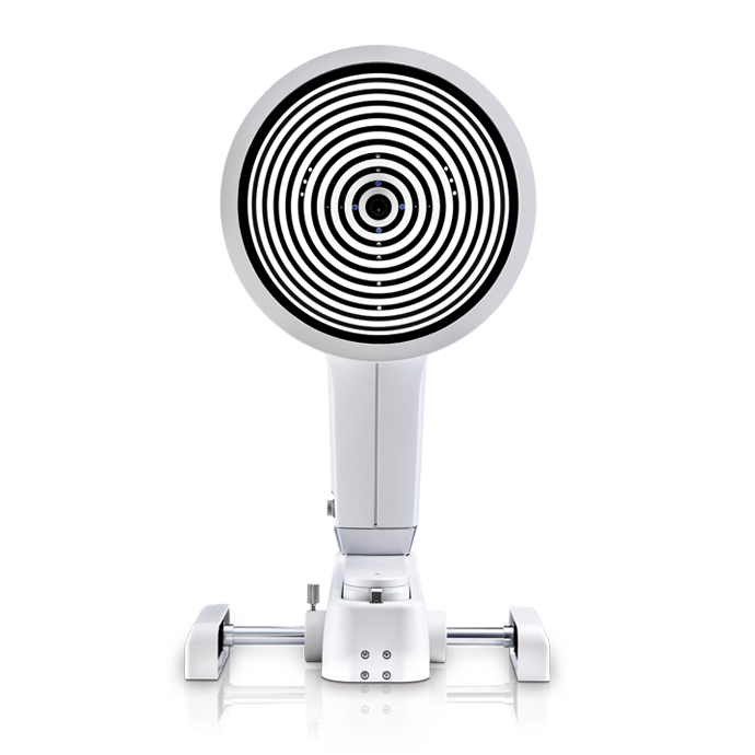
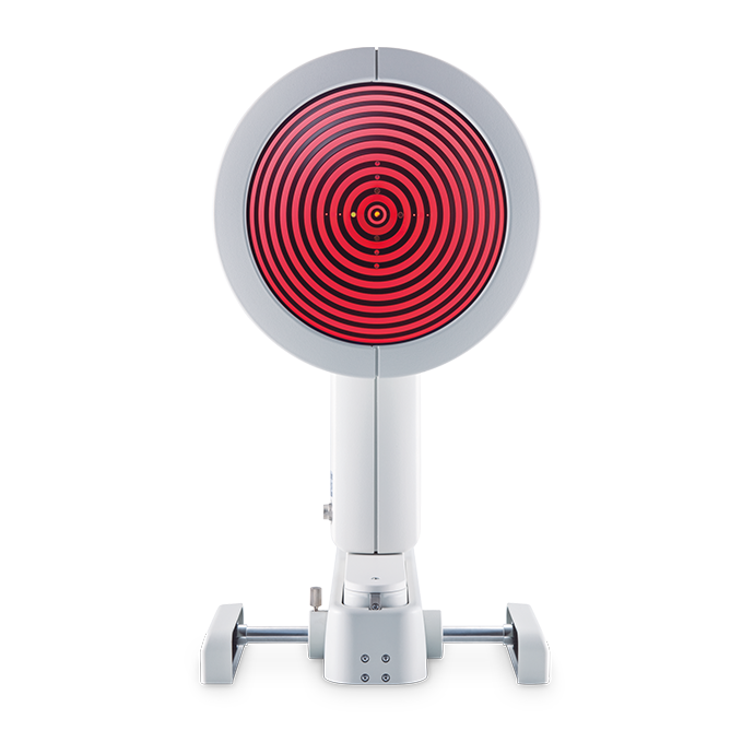
The OCULUS Keratograph® 5M
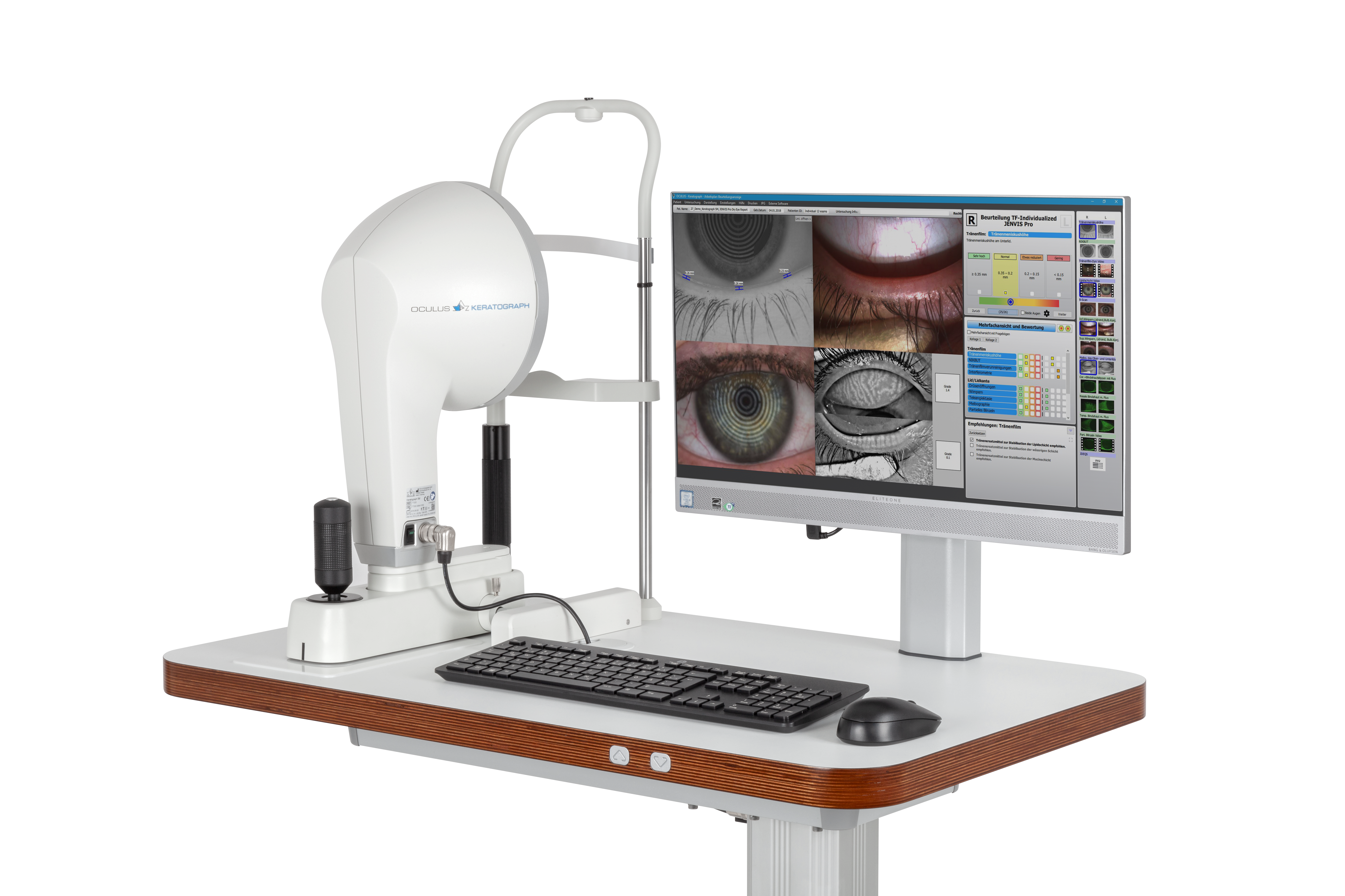
The OCULUS Keratograph® 5M is an advanced corneal topographer with a built-in real keratometer and a color camera optimized for external imaging. Unique features include examining the meibomian glands, non-invasive tear film break-up time and the tear meniscus height measurement and evaluating the lipid layer.
The keratometer: integrated in all OCULUS topography devices
The benefits of the integrated keratometer are crucial for opticians’ and optometrists’ everyday practice:
- measurements of actual corneal curvature
- exact measurements for perfect correction of contact lenses
- intuitive and simple to use
- can be processed quickly
- high-resolution images ensure clear results for customers
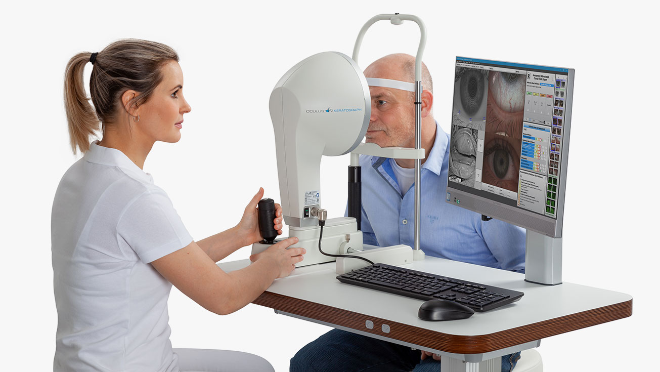

Functions
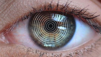
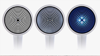
Measurements with Placido Ring Illumination
Thousands of measuring points are used to measure the whole surface of the cornea. A white ring illumination is used for this purpose. An infrared ring illumination is also provided for analysis of the tear film to prevent glare-related reflex secretion.
Measurements with Light Emitting Diodes
The perfect illumination has been integrated for every function of the Keratograph® 5M: White diodes for the tear film dynamics, blue diodes for fluo-images, infrared diodes for Meibography.
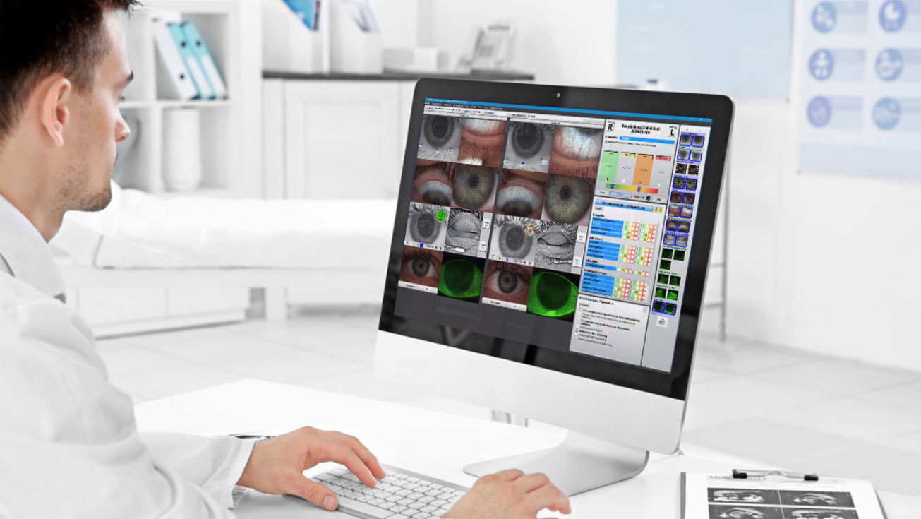
Software Highlights
Software
The heart of the Keratograph® 5M
Makes the Tear Film Visible
NIKBUT – Assessment of the Tear Film Break-Up Time
Meniscus Tear Height – Assessment of the Tear Film Quantity
Lipid Layer – Assessment of the Interference Phenomenon
TF Dynamics – Assessment of the Particle Flow
R-Scan – Assessment of the Particle Flow
Meibo-Scan – Meibography of the Top and Bottom Eyelid
Make Dry Eye Diagnosis Crystal Clear
Recording Made Easy
Excel With Your Dry Eye Diagnosis
Documentation and Patient Education
How Does That Work?
Indispensable for:
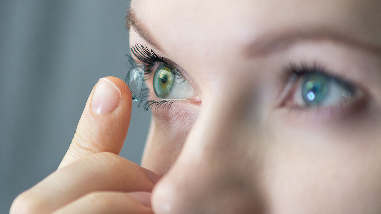
Oxygen Transmissibility of Soft Contact Lenses Made Visible
Easy-to-Understand Patient Education
Compare Different Soft Contact Lenses!
Additionally, you can enter the data from other tests, such as Osmolarity, Schirmer's Test, Phenol Red Thread Test, and more.
How Does That Work?
- Simply integrate the Imaging Software into the Keratograph® software
- Blue light-emitting diodes in the illumination beam path stimulate the fluorescein
- Yellow filters are integrated into the observation beam path
Result: Static fluorescein-images and videos can be recorded under slit lamp conditions!
- Demonstrating the fit of contact lenses
- Assessment of the static fluo-image
- Assessment of the fit of contact lenses at different pupil sizes
- Comparison of fluo-image simulations with real-time fluo-images
- Selection of the best possible contact lens
- Consultation and customer retention
An intact tear film and a good supply of oxygen to the cornea are an absolute necessity for wearing contact lenses comfortably. The OxiMap® presents a color map of the oxygen transmissibility of soft contact lenses based on the lens power – which even your customers will understand!
The Dk/t values are color-coded, whereby black represents an oxygen supply lower than that present in a closed eye. To preserve the integrity of the cornea when wearing contact lenses, minimum Dk/t values are recommended based on the length of time the contact lenses are to be worn. The OxiMap® color-coding is based on these international recommendations.
OxiMap® was developed in close cooperation with JENVIS Research and the University of Applied Sciences in Jena.
- Fitting multi-focal contact lenses
- Exact determination of the treatment area for refractive surgery
- Seamless integration into the existing Keratograph® software
- The infrared camera installed in the Keratograph delivers images of the patient’s pupil, which are used as the basis for the measurements
Different Ways of Determining Pupil Reaction:
- Examination of the pupil reaction both with and without glare
- Examination using two different glare stimulus powers
- Clear presentation of the results in graphic form: pupil changes over a period of time; minimum, maximum and mean pupil diameter, incl. standard deviation
- Comparison views possible


Title
Description
Discover How Specialty Care Experts Use the Keratograph® 5M to Benefit Their Practice & Patients
Watch our educational online seminars and testimonials from leading specialists on how the Keratograph® 5M has transformed their workflow, conversion rates, practice revenue, and more.
Increasing Profitability in Your Dry Eye Practice with the OCULUS Keratograph® 5M
Jamie Kuzniar, OD
How the Keratograph® 5M Can Elevate Your Dry Eye Clinic
Rolando Toyos, MD
A Step By Step Dry Eye Work Up, Online Seminar with Dr. Crystal Brimer, O.D.
Dr. Crystal Brimer, O.D.
Dr. Crystal Brimer, O.D.: Dry Eye Disease with Crystal Brimer [9 parts]
Dr. Crystal Brimer, O.D.
Technical Data
| Measuring range | 0.1 – 1.5 in, 9 – 99 D |
| Accuracy | ± 0.1 D |
| Reproducibility | ± 0.1 D |
| Number of rings | 22 |
| Working distance | 78-100 mm |
| Number of evaluated data points | 22,000 |
| Camera | Digital CCD camera |
| Lightsource | Placido illumination: White Placido illumination: Infrared 880 nm Fluorescein illumination: Blue 465 nm Meibography: Infrared 840 nm Tear film dynamics: White Pupillometer illumination: Infrared 880 nm |
| Dimensions (W x D x H) | 275 x 320 – 400 x 480 – 510 mm |
| Weight | Measuring head: 7.1 lbs With base: 13.5 lbs |
| Max. power consumption | 18 W |
| Voltage | 90-264 V AC |
| Frequency | 47-63 Hz |
| Recommended computer specifications | Intel® Core™ i5 (Current Generation), 1 TB HDD, 8 GB memory, Windows® 10 Pro |
| Recommended screen resolution | 1920 x 1200 pixel |
Contact us!
Request a quote or contact us if you need further information.
Request a quoteContact requestHotline
Questions?
Get in touch with us.
Tel. +1 888 284-8004
Choose your topographer
OCULUS gives you the choice. What do you need for topography in your everyday practice?
Keratograph® 5M, Keratograph® 4 or Easygraph: Whichever device you choose, OCULUS quality is assured.
|
 |
 |
|
| Keratograph® 5M | Keratograph® 4 | Easygraph | |
| Topography | |||
| Overview Presentation | ✓ | ✓ | ✓ |
| Large Color Map | ✓ | ✓ | ✓ |
| 4 Color Maps Selectable | ✓ | ✓ | ✓ |
| Camera Image | ✓ | ✓ | ✓ |
| 3D Cornea | ✓ | ✓ | ✓ |
| Fourier Analysis and Zernike Analysis | ✓ | ✓ | ✓ |
| Topographic Keratoconus Screening | ✓ | ✓ | ✓ |
| Elevation Map | ✓ | ✓ | ✓ |
| Corneal asphericity | ✓ | ✓ | ✓ |
| Lens Fitting | ✓ | ✓ | ✓ |
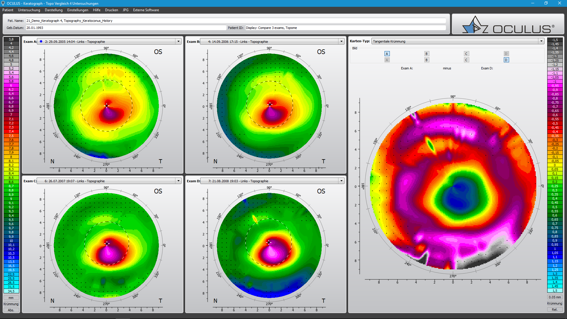
| Compare examinations | ✓ | ✓ | ✓ |
| Measurement contact lens back surface | ✓ | – | – |
| Imaging | |||
| Image and video documentation with fluo images | ✓ | ⃘ | ✓ |
| Near-Portion Height Measurement | ✓ | ✓ | – |
| Palpebral Angle Measurement | ✓ | ✓ | – |
| Manual Recording Mode | ✓ | ✓ | – |
| Crystal TEAR Report | |||
| Crystal TEAR Report (Find out the cause of dry eye disease quickly and reliably) | ⃘ | – | – |
| TF-Scan (Evaluation of lipid layer and tear film dynamics, measurement of tear meniscus height and non-invasive tear film break-up time (NIKBUT)) | ⃘ | ⃘ | – |
| R-Scan (Automatic classification of bulbar and limbal redness) | ⃘ | – | – |
| Meibo-Scan (Meibography of upper and lower eyelid) | ⃘ | – | – |
| Pupillometry | |||
| Examination of pupillary response using the pupillometer, asymmetry test and manual measuring mode | ⃘ | ⃘ | – |
| Oximap® | |||
| Raphic representation of the oxygen transmissibility (Dk/t) of soft contact lenses | ⃘ | ⃘ | ⃘ |
| Technical Data | |||
| Measuring range | 0.1 – 1.5 in, 9 – 99 D | 0.1 – 1.5 in, 9 – 99 D | 0.1 – 1.5 in, 9 – 99 D |
| Accuracy | ± 0.1 D | ± 0.1 D | ± 0.1 D |
| Reproducibility | ± 0.1 D | ± 0.1 D | ± 0.1 D |
| Number of rings | 22 | 22 | 22 |
| Working distance | 78 bis 100 mm | 80 mm | 40 mm |
| Number of evaluated data points | 22,000 | 22,000 | 22,000 |
| Camera | Digital CCD camera | Digital CCD camera | – |
| Dimensions (W x D x H) | 275 x 320 to 400 x 480 to 510 mm (10.8 x 12.6 to 15.7 x 18.9 to 20.2 in) |
275 x 320 to 400 x 490 to 517 mm (10.8 x 12.6 to 15.7 x 19.3 to 20.4 in) |
119 x 102 x 216 mm (4.7 x 4.0 x 8.5 in) |
| Weight | 7.1 lbs (Measuring head) / 13.5 lbs (with base) | 5.1 lbs (Measuring head) / 11.7 lbs (with base) | 1.6 lbs |
| Details | Details |
Legend: ✓ = available * – = not available * ⃘ = optionally available against surcharge